
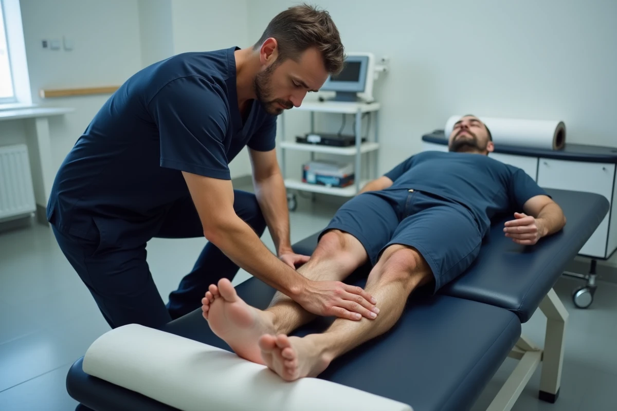
The calcaneal spur is not the direct cause of pain in the majority of cases. The calcification visible on X-ray indicates chronic tension on the enthesis of the plantar fascia, but it is the local inflammation, fasciitis, that generates the painful condition. Confusing the radiological image with the source of pain remains the most common reasoning error, leading to inappropriate therapeutic decisions.
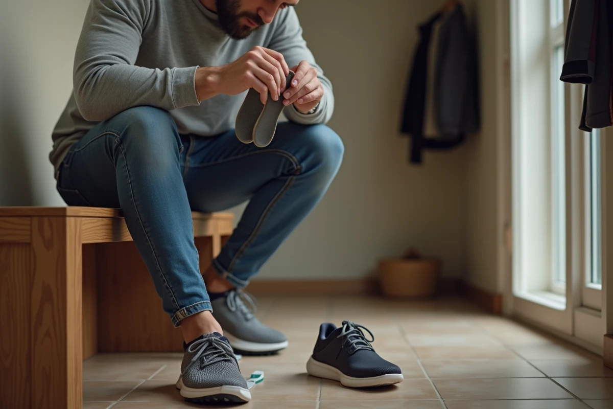
Progressive Wearing Protocol for Orthotic Insoles for Calcaneal Spur
An insole that is too corrective, too rigid, or worn all day from the start often worsens pain instead of reducing it. We regularly observe in consultations patients whose symptoms have worsened after the fitting of insoles, simply because the wearing protocol has not been followed.
Further reading : Common Mistakes When Installing Plasterboard and Solutions to Fix Them
Modern approaches recommend a distributed cushioning under the entire heel rather than localized reinforcement under the spur. The biomechanical goal is to redistribute loads across the entire foot, not to “fill” the painful area with a hard material. A soft to semi-rigid insole, with a progressive medial arch, yields better results than a highly corrective orthotic that abruptly alters pressure points.
The wearing time should increase in stages. We recommend starting with two to three hours on the first day, then adding one hour per day over a full week. This protocol allows tissues time to adapt to the new distribution of stresses. Skipping this adaptation phase can cause metatarsal pain or worsening fasciitis, which the patient mistakenly attributes to the insole itself.
Related reading : Where and how to use vacation vouchers?
To effectively treat the calcaneal spur, the insole is just one element of a comprehensive protocol that includes targeted stretching and activity load management.
Focal Shockwaves: The Therapeutic Filter Before Any Surgical Discussion
Extracorporeal focal shockwaves are now an almost mandatory step before surgery, after failure of conservative treatments over six to twelve months. This prioritization, rarely explained in mainstream content, alters the decision-making sequence for patients in therapeutic deadlock.
The distinction between radial and focal shockwaves is important. Radial waves, more accessible in clinics, disperse energy at the surface. Focal waves concentrate the beam on the insertion point of the fascia, with a deeper effect on pathological neovascularization and stimulation of tissue healing.
A standard protocol consists of three to five sessions spaced one week apart. Pain may transiently increase after the first sessions, which leads some patients to prematurely abandon treatment. This inflammatory rebound is part of the mechanism of action and does not indicate treatment failure.
Indication Criteria for Focal Shockwaves
- Persistent heel pain for more than six months despite insoles, stretching, and anti-inflammatories
- Thickening of the plantar fascia confirmed by ultrasound (above the pathological threshold)
- Absence of local contraindications (partial rupture of the fascia, infection, coagulation disorder)
- Patient functionally limited in walking or professional activity
This last point deserves attention. Recent recommendations in sports medicine now assess functional impact rather than just pain intensity. A very painful but functional patient is less likely to be a candidate for surgery than a moderately painful patient who is unable to work.
Stretching of the Plantar Fascia and Gastrocnemius: Common Technical Errors
Stretching is the foundation of conservative management, but its execution poses problems for the majority of patients. The most common mistake is stretching the calf while standing against a wall, with the knee straight, without bending the opposite knee. This position targets the gastrocnemius but ignores the soleus, whose stiffness directly contributes to the tension on the fascia.
To target the soleus, bending the knee on the stretched side is essential. We recommend holding each stretch for thirty seconds, repeated three times per side, morning and evening. Morning stretching, before placing the foot on the ground, significantly reduces pain during the first steps.
Specific Stretch for the Plantar Fascia
Sitting, cross the affected ankle over the opposite knee. Grasp the base of the toes and pull them into dorsiflexion until you feel tension under the arch. Maintaining this position for thirty seconds before each rise reduces the stiffness of the fascia accumulated overnight. This exercise directly targets the aponeurosis, where calf stretches only act indirectly.
Another common error: rolling the foot on a hard ball (golf ball, tennis ball) with excessive pressure. This action compresses the inflammatory tissues instead of mobilizing them. A cold water bottle rolled under the arch combines gentle mobilization with a local anti-inflammatory effect, with less risk of worsening the enthesopathy.
Corticosteroid Injections and Surgery for Calcaneal Spur: When to Consider Them
Corticosteroid injections still have a place in the therapeutic arsenal, but their indications have narrowed. More than two injections per year at the same site increase the risk of rupture of the plantar fascia. Relief can be spectacular in the short term, prompting repetition of the procedure, but cumulative tissue weakening makes this strategy counterproductive beyond two to three injections.
Surgery (partial fasciotomy, spur resection) is only considered after documented failure of the entire conservative protocol, including shockwaves, over a minimum duration of twelve months. Indications are based on objectively assessed functional impact, not on the size of the spur on X-ray.
- Endoscopic fasciotomy: partial section of the fascia at its calcaneal insertion, faster recovery than open surgery
- Isolated spur resection: rarely performed alone, as calcification is not the source of pain
- Baxter’s nerve release: to be considered if a canal syndrome is associated, often underdiagnosed
The main trap remains focusing on the radiological image. A large spur in an asymptomatic patient does not justify any intervention. Conversely, severe fasciitis with a minimal or absent spur requires the same therapeutic rigor. Treatment targets inflammation and mechanical overload, not calcification.